| Lines of evidence from various areas of research suggest a biological
basis of ADHD:
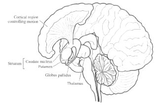
Damage to prefrontal cortex associated with ADHD-like
behavior; ADHD deficits on neuropsychological tests associated w.
prefrontal lobe function; autonomic under-arousal (GSR, heart rate,
etc); decreased blood flow to prefrontal regions and striatum
(globus pallidus, caudate nucleus, putamen); diminished metabolic
activity in left anterior frontal correlates with severity of ADHD
symptoms.
fMRI Findings: differences in size of temporal lobe (speech areas); corpus
callosum; prefrontal cortex, and basal ganglia seen in ADHD versus
normals. Differences in metabolic rate as indicated by blood flow during
tasks requiring attention also seen.
Stimulants (amphetamines and related compounds) cause dopaminergic
neurons that project to (among other structures) the basal ganglia to
secrete more dopamine. Basal ganglia structures have receptors for
dopamine. Basal ganglia are involved in control of learned motor
sequences, repetitive behavior, and operant (reward-based) learning and
performance. Stimulants reduce symptoms of ADHD, presumably by
increasing availability of dopamine.
|